Anatomy Arteries In Neck : blood pressure - Which vessel could a pumping vessel in ... / The common carotid artery is a primary source of oxygenated blood to the head and neck.
byAdmin-
0
Anatomy Arteries In Neck : blood pressure - Which vessel could a pumping vessel in ... / The common carotid artery is a primary source of oxygenated blood to the head and neck.. Note the feathery network of blood vessels in the left and right lungs (next to the heart). Anatomy atlases, the anatomy atlases logo, and a digital library of anatomy information are all trademarks of michael p. Illustration of the arterial system in the human body, shown in a standing figure. Cca typically divides at the level of c3 or c4 vertebral. Instant anatomy is a specialised web site for you to learn all about human anatomy of the body with diagrams, podcasts and revision questions.
The left and right carotids, and the left and right vertebral arteries. Vagus nerve or sympathetic plexus: The arteries and veins of the face and neck were labeled focusing on large trunks emerging from the external carotid artery or draining into the. 512 anatomical structures were dynamically labeled, and some structures have been redesigned or enhanced with a graphic tablet for better readability. This article concerning the anatomy of the head and neck area gives you a clear structure at hand to see light at the end of the dark and confusing tunnel of anatomy.
Arteries of the Neck and Head - Human Anatomy from medicalterms.info Some important structures contained in or passing through the neck include the seven cervical vertebrae and enclosed spinal cord, the jugular veins and carotid arteries, part of the esophagus, the larynx. Muscle head anatomy vocal organ diagram female neck anatomy neck wireframe head neck human anatomy head artery anatomy face pharynx vector neck degree head anatomy 3d. Note the feathery network of blood vessels in the left and right lungs (next to the heart). The cca courses superiorly in the neck, anteromedial to the jugular vein and alongside the vagus nerve. There are many possible anatomical configurations of the aortic arch and its branches. The head and neck receives the majority of its blood supply through the carotid and vertebral arteries. The common carotid artery is a primary source of oxygenated blood to the head and neck. The easiest spot is where it joins your head, just under the corner of the mandible.
And anterior deep temporal a.
The mass is not in between the. The information contained in anatomy atlases is not a substitute for the medical care and advice of your physician. Anatomy of the head and neck. The infrahyoid neck is divided into 5 major anatomical compartments or spaces by the various layers of the cervical fascia (2) carotid artery and internal jugular vein: There may be variations in treatment that. The carotid arteries extend out from the aorta artery, which transports blood out of the heart and is the body's largest artery. The arteries and veins of the face and neck were labeled focusing on large trunks emerging from the external carotid artery or draining into the. All major arteries of the neck originate from the aortic arch via three main vessels: See more ideas about arteries, head and neck, anatomy and physiology. Thus the vascular territory supplied by the striate arteries is particularly susceptible to lacunar infarcts. There are many possible anatomical configurations of the aortic arch and its branches. The lingual artery, which supplies the tongue as well as the oral floor, is a major branch of the external carotid artery. Vagus nerve or sympathetic plexus:
Head and neck anatomy quiz 2: Illustration of the arterial system in the human body, shown in a standing figure. See more ideas about arteries, head and neck, anatomy and physiology. The carotid arteries extend out from the aorta artery, which transports blood out of the heart and is the body's largest artery. The external carotid artery stretches upwards from the level of upper border of the lamina of the thyroid cartilage to a stage supporting the neck of the mandible, where it ends in the substance of the parotid gland by splitting.
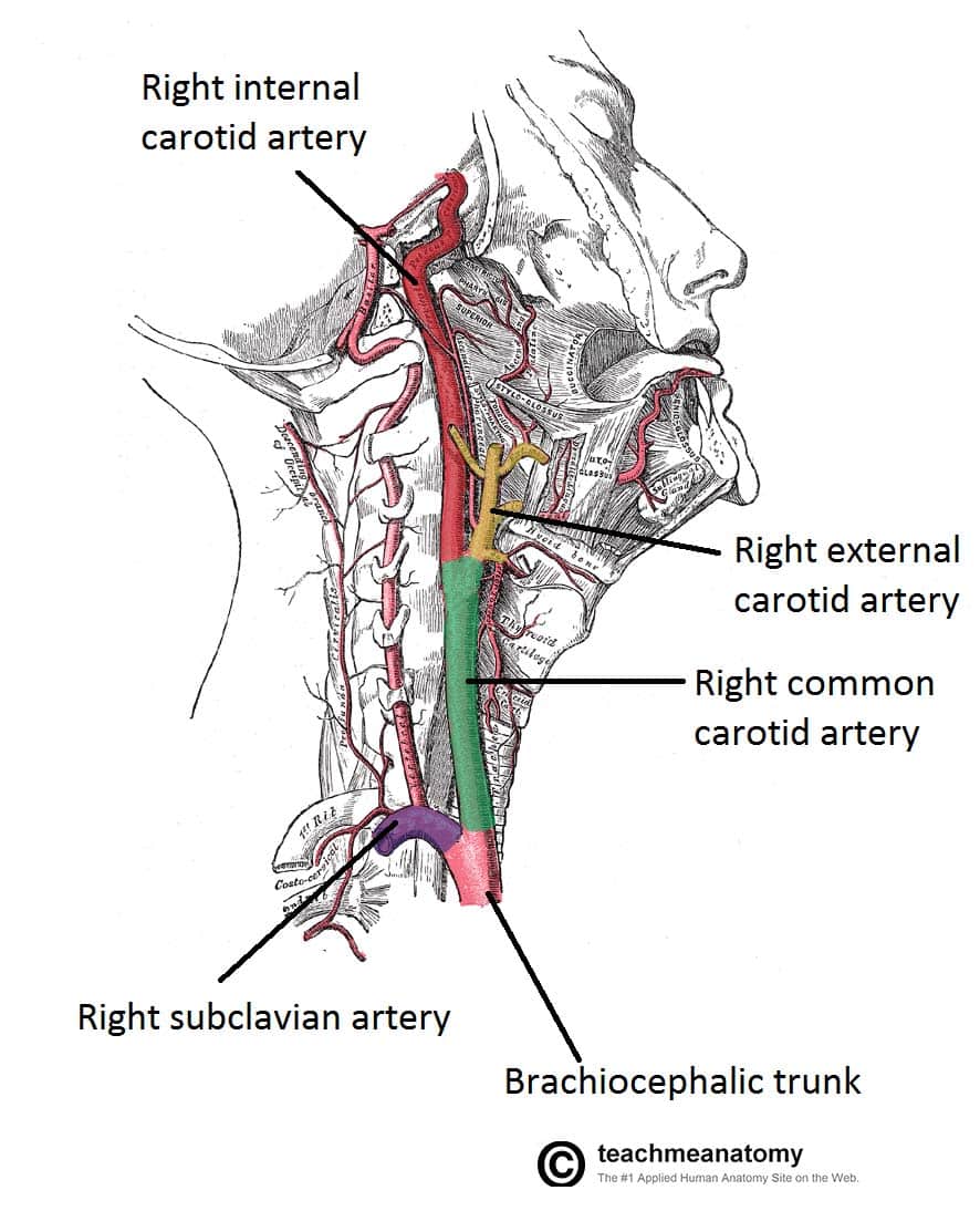
Blood Vessels and Lymphatics of the Head and Neck ... from teachmeanatomy.info Part of the occipital and parts of the transverse cervical and suprascapular arteries are also found in the occipital triangle. The information contained in anatomy atlases is not a substitute for the medical care and advice of your physician. The position of the branched carotid arteries is where a person can feel the pulse in their neck, just under the jaw. Anatomy ▶ head and neck ▶ arteries ▶ common carotid arteries. The right and left subclavian arteries give rise to the thyrocervical trunk. There may be variations in treatment that. The external carotid artery stretches upwards from the level of upper border of the lamina of the thyroid cartilage to a stage supporting the neck of the mandible, where it ends in the substance of the parotid gland by splitting. The anatomy of neck arteries, normal variations, and anastomoses between different arteries is discussed in this chapter.
The left and right carotids, and the left and right vertebral arteries.
There may be variations in treatment that. The left and right carotids, and the left and right vertebral arteries. It is a branchless artery that travels up the neck lateral to the trachae and larynx, to the upper boarder of the thyroid cartilage. Some important structures contained in or passing through the neck include the seven cervical vertebrae and enclosed spinal cord, the jugular veins and carotid arteries, part of the esophagus, the larynx. An artery is an elastic blood vessel that transports blood away from the heart. Cca typically divides at the level of c3 or c4 vertebral. And anterior deep temporal a. The anatomy of neck arteries, normal variations, and anastomoses between different arteries is discussed in this chapter. In this video, we discuss arterial supply to the head and neck including the aorta, brachiocephalic trunk, common carotid, and subclavian artery. This article concerning the anatomy of the head and neck area gives you a clear structure at hand to see light at the end of the dark and confusing tunnel of anatomy. Learn about the types of arteries and how they function. See more ideas about arteries, head and neck, anatomy and physiology. In radiology, the 'head and neck' refers to all the anatomical structures in this region excluding the central nervous system, that is, the brain and spinal co.
The infrahyoid neck is divided into 5 major anatomical compartments or spaces by the various layers of the cervical fascia (2) carotid artery and internal jugular vein: Neck, in land vertebrates, the portion of the body joining the head to the shoulders and chest. This section of the website will explain large and minute details of arterial anatomy of neck. Illustration of the arterial system in the human body, shown in a standing figure. And anterior deep temporal a.
Arteries of the Face and Neck | Plastic Surgery Key from plasticsurgerykey.com There are an additional eight major divisionstrusted. These arteries do not have significant collateral circulation; The anatomy of neck arteries, normal variations, and anastomoses between different arteries is discussed in this chapter. The carotids reside beneath the skin on either side, and the pulse can be felt easily with your hand. It is a branchless artery that travels up the neck lateral to the trachae and larynx, to the upper boarder of the thyroid cartilage. Through its external carotid branch, it supplies the face, scalp, tongue, upper and lower teeth, gums, sinus, external and middle ear, pharynx and larynx in the throat, as well as the thyroid. 37.11 arteries of the neck left lateral view. Head and neck anatomy is important when considering pathology affecting the same area.
In radiology, the 'head and neck' refers to all the anatomical structures in this region excluding the central nervous system, that is, the brain and spinal co.
The anatomy of neck arteries, normal variations, and anastomoses between different arteries is discussed in this chapter. It is a branchless artery that travels up the neck lateral to the trachae and larynx, to the upper boarder of the thyroid cartilage. There are many possible anatomical configurations of the aortic arch and its branches. Arteria carotis interna) is located in the inner side of the neck in contrast to the external carotid artery. These arteries do not have significant collateral circulation; The position of the branched carotid arteries is where a person can feel the pulse in their neck, just under the jaw. An artery is an elastic blood vessel that transports blood away from the heart. Vagus nerve or sympathetic plexus: The anatomy of neck arteries, normal variations, and anastomoses between different arteries is discussed in this chapter. A publicly available article also appearing in pubmed about anatomy, head and neck, striate arteries. The external carotid artery stretches upwards from the level of upper border of the lamina of the thyroid cartilage to a stage supporting the neck of the mandible, where it ends in the substance of the parotid gland by splitting. The neck is supplied by arteries other than the carotids. There are an additional eight major divisionstrusted.
Form the arch of the aorta behind the centre of the manubrium (it enters the neck behind the surface anatomy is represented by a line extending from the sternoclavicular joint to the upper border of the thyroid cartilage arteries in neck. Learn about the types of arteries and how they function.